Presents you biological research related topics such as biotechnology, biochemistry, microbiology etc. explained in a simple and easy to understand form. Let's make science easy !!!
All they do is sense along with vestiblo cochlear that bears only 8 notes
3&4 I call you motor because I am driving the truck clear
Roll no 5. Trigemi is a mixed child
Who was abducted at 6’o clock in a car by 7 known faces mixed in the crowd
My 9th grade friend Glossy pharynx mixed with the crowd on 10th at Las Vegas who have 11 accessorized cars but their 12th car is less glossy
One of the most dreaded tasks for anatomy students is learning the names of the 12 cranial nerves. I, like many other students before me, despised memorising them. Instead, I tired looking for mnemonics to remember the cranial nerves. I will list down below a few mnemonics that I particularly useful. However, in my search for good mnemonics, I ended up creating my own. The poem above was my method of recalling the names of the 12 cranial nerves during my student days.
The source of a protein is generally tissue or microbial cells. The first step in any protein purification procedure is to break open these cells, releasing their proteins into a solution called a crude extract. If necessary, differential centrifugation can be used to prepare sub-cellular fractions or to isolate specific organelles.
Commonly, the extract is subjected to treatments that separate the proteins into different fractions based on a property such as size or charge, a process referred to as fractionation. Early fractionation steps in a purification utilize differences in protein solubility, which is a complex function of pH, temperature, salt concentration, and other factors.
The solubility of proteins is lowered in the presence of some salts, an effect called ‘salting out’. The addition of certain salts in the right amount can selectively precipitate some proteins, while others remain in solution. Ammonium sulfate ((NH4)2SO4) is particularly effective and is often used to salt out proteins. The proteins thus precipitated are removed from those remaining in solution by low-speed centrifugation.
Dialysis is a procedure that separates proteins from small solutes by taking advantage of the proteins’ larger size. The partially purified extract is placed in a bag or tube made of a semipermeable membrane. When this is suspended in a much larger volume of buffered solution of appropriate ionic strength, the membrane allows the exchange of salt and buffer but not proteins. Thus dialysis retains large proteins within the membranous bag or tube while allowing the concentration of other solutes in the protein preparation to change until they come into equilibrium with the solution outside the membrane. Dialysis might be used, for example, to remove ammonium sulfate from the protein preparation.
Column Chromatography: A porous solid material with appropriate chemical properties (the stationary phase) is held in a column, and a buffered solution (the mobile phase) migrates through it. The protein, dissolved in the same buffered solution that was used to establish the mobile phase, is layered on the top of the column and allowed to percolate into the solid matrix by the addition of the buffer solution on top. The protein solution forms a band within the mobile phase that is initially the depth of the protein solution applied to the column. As proteins migrate through the column, they are retarded to different degrees by their different interactions with the matrix material. Individual types of proteins gradually separate from each other, forming bands within the broader protein band.
Separation improves (i.e., resolution increases) as the length of the column increases. However, each individual protein band also broadens with time due to diffusional spreading, a process that decreases resolution.
Three types of column chromatographic methods used in protein purification.
(a) Ion-exchange chromatography exploits differences in the sign and magnitude of the net electric charges of proteins at a given pH.
(b) Size-exclusion chromatography, also called gel filtration, separates proteins according to size.
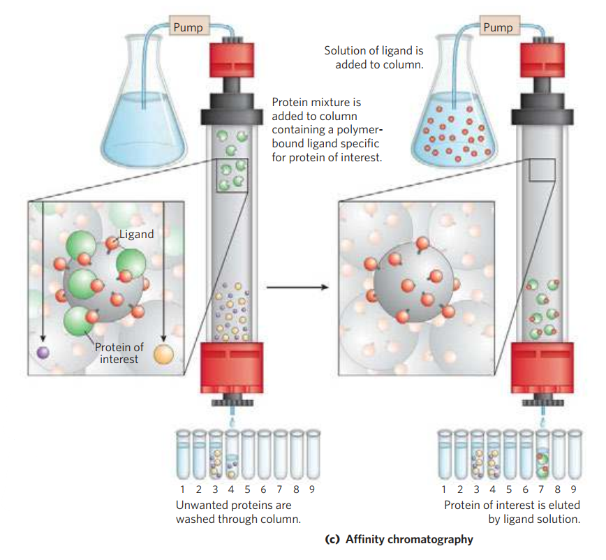
(c) Affinity chromatography separates proteins by their binding specificities.
Ion-Exchange Chromatography: The column matrix is a synthetic polymer (resin) containing bound charged groups; those with bound anionic groups are called cation exchangers, and those with bound cationic groups are called anion exchangers. The affinity of each protein for the charged groups on the column is affected by the pH (which determines the ionization state of the molecule) and the concentration of competing free salt ions in the surrounding solution. Separation can be optimized by gradually changing the pH and/or salt concentration of the mobile phase so as to create a pH or salt gradient. In cation-exchange chromatography, the solid matrix has negatively charged groups. In the mobile phase, proteins with a net positive charge migrate through the matrix more slowly than those with a net negative charge, because the migration of the former is retarded more by interaction with the stationary phase.
Size-Exclusion Chromatography: In this method, large proteins emerge from the column sooner than small ones. The solid phase consists of cross-linked polymer beads with engineered pores or cavities of a particular size. Large proteins cannot enter the cavities and so take a shorter (and more rapid) path through the column, around the beads. Small proteins enter the cavities and are slowed by their more labyrinthine path through the column. Size-exclusion chromatography can also be used to approximate the size of a protein being purified similar to gel electrophoresis.
Affinity Chromatography: The beads in the column have a covalently attached chemical group called a ligand—a group or molecule that binds to a macromolecule such as a protein. When a protein mixture is added to the column, any protein with affinity for this ligand binds to the beads, and its migration through the matrix is retarded. For example, if the biological function of a protein involves binding to ATP, then attaching a molecule that resembles ATP to the beads in the column creates an affinity matrix that can help purify the protein. As the protein solution moves through the column, ATP-binding proteins (including the protein of interest) bind to the matrix. After proteins that do not bind are washed through the column, the bound protein is eluted by a solution containing either a high concentration of salt or free ligand—in this case, ATP or an analog of ATP. Salt weakens the binding of the protein to the immobilized ligand, interfering with ionic interactions. Free ligand competes with the ligand attached to the beads, releasing the protein from the matrix; the protein product that elutes from the column is often bound to the ligand used to elute it.
Chromatographic methods are typically enhanced by the use of HPLC, or high-performance liquid chromatography. HPLC makes use of high-pressure pumps that speed the movement of the protein molecules down the column, as well as higher-quality chromatographic materials that can withstand the crushing force of the pressurized flow. By reducing the transit time on the column, HPLC can limit diffusional spreading of protein bands and thus greatly improve resolution.
Gel Electrophoresis is a technique used to separate DNA, RNA or protein molecules based on their size, electrical charge and conformation using electric field at a definite pH.
It is based on the principle that charged molecules when placed in an electric field, tend to migrate towards the electrode of the opposite charge.
Many important biological molecules, such as amino acids, peptides, proteins, nucleotides and nucleic acids, possess ionisable groups and, therefore, at any given pH, exist in solution as electrically charged species, either cations (positively charged) or anions (negatively charged).
Gel Electrophoresis makes use of porous gels that act as a molecular sieve separating bigger molecules from the smaller ones.
The rate at which each molecule travels through the gel is called its electrophoretic mobility and its determined mainly by its net charge, size & shape.
Strongly charged molecules move faster than weakly charged ones.
Smaller molecules move faster than larger ones.
Molecules with highly coiled structures move faster than uncoiled ones.
Nature of charge:
Under the influence of an electric field these charged particles will migrate either to cathode or anode depending on the nature of their net charge.
Nucleic acids which are negatively charged, due to the presence of the sugar-phosphate backbone, migrate toward the anode which is positively charged.
Amino acids that make up proteins may be positive, negative, neutral, or polar in nature depending on the functional groups present in the amino acid. The charges of amino acids together give a protein its overall charge.
Thus, when researchers want to separate proteins using gel electrophoresis, they must first mix the proteins with a detergent called sodium dodecyl sulfate (SDS). This treatment makes the proteins unfold into a linear shape and coats them with a negative charge, which allows them to migrate toward the positive end of the gel and be separated.
Voltage:
When a potential difference (voltage) is applied across the electrodes, it generates a potential gradient (E), which is the applied voltage (V) divided by the distance “d” between the two electrodes i.e.
E = V/d
When this potential gradient ‘E’ is applied, the force as the molecule bearing a charge of ‘Q’ is
F = E x Q
Unit of Force (F) is Newtons & Unit of Charge (Q) is coulombs
It is this force that drives the molecule towards the electrodes.
Frictional force:
There is also a frictional force that retards the movement of this charged molecule.
This frictional force depends on :
Hydrodynamic size of the molecule
Shape of the molecule
Pore size of the medium in which the electrophoresis is taking place
Viscosity of the buffer.
The velocity ‘v’ of the charged molecule is an electric field is therefore given by the equation :
V = (E x Q)/f =(V x Q)/(d x f)
where ‘f’= frictional coefficient
Electrophoretic mobility:
Electrophoretic mobility ( u ) of an ion is represented as the ratio of the velocity of the ion and the field strength. i.e.
u =V/E
When a p.d. is applied, the molecule with different overall charges will begin to separate owing to their different electrophoretic mobility.
Even the molecule with similar charges will begin to separate if they have different molecular sizes, since they will experience different frictional forces.
Current:
According to Ohm’s law:
V/I=R
Therefore, it appears that it is possible to accelerate an electrophoretic separation by increasing the applied voltage, which ultimately results in corresponding increase in the current flowing.
The distance migrated by the ions will be proportional to both current and time.
Heat:
One of the major problems for most forms of electrophoresis, that is the generation of heat.
During electrophoresis, the power ( W ) generated in one supporting medium is given by
W= I2R
Most of the power generated is dissipated as heat.
The following effects are seen on heating of the electrophoretic medium has :
An increased rate of diffusion of sample and buffer ions which leads to the broadening of the separated samples.
Formation of convention currents, which leads to mixing of separated samples.
Thermal instability of samples that are sensitive to heat.
A decrease of buffer viscosity and hence reduction in the resistance of the medium.
If a constant voltage is applied, the current increases during electrophoresis owing to the decrease in resistance and this rise in current increases the heat output still further.
For these reasons, often a stabilized power supply is used, which provides constant power and thus eliminates fluctuations in heating.
Constant heat generation is however a problem. For which the electrophoresis is run at very low power (low current) to overcome any heating problems, but this can lead to poor separation as a result of the increased amount of diffusion due to long separation time.
Compromise condition have to be found out with reasonable power settings, to give acceptable separation time and an appropriate cooling system, to remove liberated heat. While such system works fairly well, the effect of heating are not always totally eliminated.
Electroendosmosis:
Electroendosmosis occurs due to the presence of charged groups on the surface of the support medium.
For instance, paper has some carboxyl group present, agarose contains sulfate groups depending on the purity grade and the surface of glass walls used in capillary electrophoresis contains silanol (Si-OH) groups.
These groups, at appropriate pH, will ionize, generating charged sites.
It is these charges that generate electroendosmosis.
In case of capillary electrophoresis, the ionized sianol groups creates an electrical double layer, or a region of charge separation, at the capillary wall/electrolytic interface.
When voltage is applied cations in the electrolyte near the capillary walls migrate towards the cathode, pulling electrolyte solution with them.
This creates a net electro osmotic flow towards cathode.
I am sorry I haven’t posted for a while. You see I live in India and over here the Covid-19 situation is pretty bad. I can’t bring myself to post about happy and interesting stuff when I am constantly thinking about the point of life. So many people are dying on the streets, not even in the hospitals. Whether I watch the news, or scroll through social or even just check my mail all I see are people struggling, desperately asking for help.
The main thought that has been going on in my mind these days, is there no value of human life? I guess this should not be a new realization (at least for me), but still, this thought haunts me. Every time I see or hear about a person dying, or about the manner a patient or dead body was treated, I feel like somebody is holding my heart and wringing it hard in my chest, draining out the blood in it. The dark reality of our healthcare system has never been unknown to me but for the most part, it’s something that is not discussed in the media. Making the problems less visible and therefore allowing the public to ignore them. But here are a few stories about my experience with the healthcare system in the past decade that I want to share with you guys.
My maternal grandmother died 9 years in the month of May, we still don’t have her death certificate. Because of bureaucratic issues, we were never got a document registering her death in the records of the municipal corporation. Even 9 years ago, when she was sick we struggled to get a bed in the medical college. Even a decade, way way before corona hit us, accessing public healthcare was very difficult.
My paternal grandmother died in 2016, people from the municipal corporation came to collect the dead body after 7 hrs of her passing away. We had to buy a block of ice (which arrived in less than half an hour) to keep the body from smelling. Today journalists think it is a story worth covering, why was it not worth covering 5 years ago? Why did you ignore it back then? Not sensational enough?
When both my paternal grandparents died (because of our previous experience), the first thing we did was not mourn their deaths but go stand in the line for the death certificates. For my grandfather who passed away in 2019, I had to stand for 5 hours to get the certificate.
Do you realize how dehumanizing these situations felt? We didn’t have the privilege to be emotional about the experience of a loved one passing away, we had to think about logistics. This has always been the case with the health system, we just ignored this. Look where this has got us! But do you know what my biggest fear is? Right now the media is covering this, because of covid. What happens when there is no more covid? Do we just go back to ignoring this problem? This has happened in the past with diseases like AIDS, Polio, Japanese encephalitis, etc. Every time there is a new disease, the government makes many schemes and programs to counter that disease. But the government never attempts to improve the country’s overall healthcare system. So every time we have a new one we run into the same problems.
We need a system that assures all citizens access to affordable healthcare, we need better funding in healthcare and medical research, WE NEED RIGHT TO HEALTH TO BE A FUNDAMENTAL RIGHT OF ALL INDIAN CITIZENS.